
When you suddenly see a storm of black spots or flashes of light in your vision, it’s easy to brush it off as eye strain or aging. But if you’re experiencing these symptoms along with a shadow creeping across your sight, retinal detachment could be happening-and every minute counts.
Retinal detachment isn’t just a blurry vision issue. It’s a medical emergency where the thin layer of tissue at the back of your eye, called the retina, peels away from its blood supply. Without quick treatment, the photoreceptor cells that turn light into signals your brain understands start dying. Permanent vision loss can happen in hours, not days. The good news? If caught early, surgery can save your sight. The bad news? Most people wait too long.
What Are the Warning Signs You Can’t Ignore?
Retinal detachment doesn’t sneak up quietly. It screams with six clear symptoms that show up fast and get worse quickly.
- Sudden increase in floaters: Not just one or two. Patients describe it as a shower of dark specks, strings, or cobwebs filling their vision. The National Eye Institute says it’s often a dramatic change-something you’ve never seen before.
- Flashes of light: These aren’t the kind you get when you rub your eyes. They’re brief, bright bursts of light, usually in your peripheral vision, like camera flashes or lightning strikes in the corner of your eye.
- A dark curtain or shadow: This is the red flag. It starts in your side vision and slowly moves toward the center, like a curtain being pulled across your sight. NYU Langone Health calls this the most urgent sign.
- Sudden blurry or distorted vision: Things look wavy, warped, or out of focus-even if you have perfect glasses or contacts. Cleveland Clinic found this happens in nearly 7 out of 10 cases.
- Loss of peripheral vision: You might notice you can’t see things out of the corner of your eye. The Retina Research Foundation reports this occurs in 73% of cases.
- Color changes: Colors may seem washed out or dull, especially if the macula (the center of your retina) is involved. This is a sign the detachment is spreading.
If you have even one of these, especially a shadow or sudden flashes, don’t wait. Don’t call your primary care doctor. Don’t wait until tomorrow. Go to an eye specialist today.
How Doctors Diagnose It-Fast
There’s no home test for retinal detachment. You need a specialist with the right tools. The gold standard is a dilated fundus exam. That means drops widen your pupil so the doctor can see the back of your eye with an indirect ophthalmoscope and a special lens.
If your eye is cloudy from cataracts or bleeding, they’ll use B-scan ultrasound. It’s a quick, painless scan that creates a picture of your retina using sound waves. For detailed images, they’ll use optical coherence tomography (OCT), which maps the layers of your retina in microns.
General ophthalmologists miss about 22% of early detachments. Retinal specialists get it right 95% of the time. That’s why if you’re at risk or have symptoms, you need to see a vitreoretinal specialist-not just any eye doctor.
The Three Main Surgeries-And Which One Is Right for You
There’s no one-size-fits-all fix. The right surgery depends on where the tear is, how big it is, whether the macula is still attached, and your overall eye health.
1. Pneumatic Retinopexy
This is the least invasive option. A gas bubble is injected into your eye. You then position your head so the bubble floats up and presses against the detached area. The doctor seals the tear with laser or freezing therapy. It works best for small, upper retinal breaks in people who still have their natural lens.
Success rate: 70-80%. But if the tear is on the bottom of your eye, this won’t work. And you’ll need to stay in a face-down or side-lying position for 50% of the day for 7-10 days. That’s hard. Many patients report neck pain, fatigue, and difficulty reading or eating.
2. Scleral Buckling
A silicone band is stitched around the outside of your eye, gently pushing the wall inward to meet the detached retina. It’s like putting a belt around your eyeball to hold things in place. This method doesn’t require gas bubbles, so positioning isn’t as strict.
Success rate: 85-90%. Great for younger patients with lattice degeneration or large tears. But it often causes nearsightedness (1.5-2.0 diopters) and can lead to double vision in 5-8% of cases. It’s also less common now, mostly used when the vitreous is still thick and healthy.
3. Vitrectomy
This is the most common surgery today-used in about 65% of cases. The surgeon removes the gel-like vitreous inside your eye and replaces it with a gas or silicone oil bubble. They then seal the tear with laser or freezing. It’s the go-to for complex cases: giant tears, scar tissue, or when the macula is already detached.
Success rate: 90-95%. But there’s a trade-off. If you still have your natural lens, you’ll likely develop a cataract within two years-in 70% of cases. You’ll also need to position your head carefully if gas is used. Recovery takes longer, but it’s the best shot at saving central vision.
Time Is Vision-Why Every Hour Matters
Dr. Carl Regillo at Wills Eye Hospital says, “Every hour counts.” That’s not a slogan. It’s data.
A 2022 study in the Journal of VitreoRetinal Diseases found that if surgery happens within 24 hours of symptoms, the chance of full anatomical repair is 90%. If you wait 72 hours, your chance of regaining 20/40 vision drops from 75% to 35%.
One Reddit user, "VisionWarrior22," ignored floaters for three days. By the time they got help, the shadow had reached their center vision. Their final vision was 20/100-far worse than the 20/25 they could’ve had.
Most patients delay because they think it’s just “eye strain” or “old age.” The American Society of Cataract and Refractive Surgery found 63% of patients were misdiagnosed by non-eye doctors. That delay averages 48 hours. That’s two full days of photoreceptors dying.
If your macula is still attached, you’re in the best possible window. If it’s already detached, surgery can still help-but you’ll likely have some permanent blurriness or distortion. That’s why acting fast isn’t just smart-it’s essential.
Who’s at Highest Risk?
Retinal detachment affects about 1 in 10,000 people each year. But certain groups are far more vulnerable:
- People with severe myopia (nearsightedness): If your prescription is worse than -5.00, your risk jumps to 167 in 10,000.
- Post-cataract surgery patients: Your risk increases by 0.5-2% after lens removal.
- Those with lattice degeneration: This thinning of the retina affects 1 in 10 people and carries a 1% lifetime risk of detachment.
- People over 40: Incidence doubles after age 40.
- Those with a family history: If a close relative had it, your risk is higher.
Even if you don’t fit these categories, don’t ignore sudden changes. One in five cases happen in people with no known risk factors.
What to Expect After Surgery
Surgery isn’t the end-it’s the start of recovery.
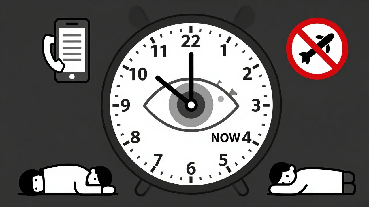
If you had a gas bubble, you’ll need to keep your head in a specific position for 7-10 days. That means sleeping on your side, eating with your head tilted, and avoiding flying or going to high altitudes until the gas is gone. You can’t fly for weeks-changes in cabin pressure can make the gas expand and damage your eye.
Side effects are common: blurry vision for weeks, redness, mild pain, and floaters from the gas. Most improve over time. But complications happen: cataracts (70%), high eye pressure (25%), and recurrence (5-15%).
Many patients need help at home. The Retina Society found 38% required home health visits for positioning help or daily check-ins. Don’t try to power through alone.
What’s New in Retinal Surgery?
Technology is getting better. In January 2023, the FDA approved the EVA Platform-a 27-gauge vitrectomy system that uses smaller incisions, less trauma, and faster healing.
Intraoperative OCT is now used in top centers. It gives surgeons real-time, high-res images of the retina during surgery, helping them remove scar tissue more completely. One study showed a 15% improvement in results.
Looking ahead, bioengineered retinal patches are in early trials. Gene therapies for inherited conditions may one day prevent detachment before it starts. But for now, the best tool is still early detection.
What You Can Do Right Now
Don’t wait for a routine eye exam. If you notice sudden flashes, floaters, or a shadow:
- Call a retina specialist immediately. Don’t wait for your regular eye doctor’s next appointment.
- If you can’t get in same-day, go to an emergency eye clinic or hospital with an ophthalmology department.
- Write down exactly when symptoms started and what you saw. This helps the doctor assess urgency.
- Don’t drive yourself if your vision is blurry. Have someone take you.
There’s no way to prevent retinal detachment if you’re at risk-but you can prevent vision loss by acting fast. Your eyes don’t have a reset button. Once the retina dies, it doesn’t come back.
Know the signs. Trust your vision. Act now.

Jason Silva
December 22, 2025 AT 04:48mukesh matav
December 22, 2025 AT 10:26Peggy Adams
December 24, 2025 AT 00:44Theo Newbold
December 24, 2025 AT 03:34Jay lawch
December 25, 2025 AT 08:39Christina Weber
December 26, 2025 AT 10:35Michael Ochieng
December 28, 2025 AT 04:32Dan Adkins
December 29, 2025 AT 05:34